Researchers from the Worcester Polytechnic Institute have created a first-of-its-kind system that combines MRI imaging and robotics through a special motor-control technology – and the results could mean a more-effective way to treat conditions such as Parkinson’s, severe depression and obsessive-compulsive disorder.
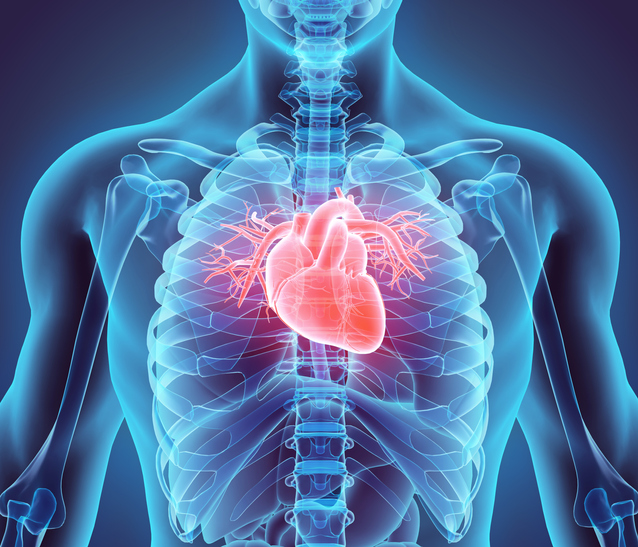
Stereotactic neurosurgery enables surgeons to target and treat diseases affecting deep structures of the brain using deep brain stimulation (DBS). But the current system is time-consuming and challenging because surgeons can’t see what’s happening inside the brain during operation. This renders them nearly blind to complications such as brain shift and target inaccuracy.
These shortcomings motivated researchers led by Professor Greg Fischer to develop an MRI-guided robotic system for DBS procedures. Published in IEEE Transactions on Biomedical Engineering, the robotic system allows a DBS surgery to take place inside the bore of an MRI, taking advantage of real-time MRI images. The system allows the neurosurgeon to precisely place electrodes intended to electrically stimulate different parts of brain structure. The neurosurgeon can use MRI imaging as a guide, observing what’s happening inside the brain during the procedure. This advancement improves target accuracy and enhances safety for the patient, as well as reduces surgery time.

- Professor Greg Fischer and graduate research student Hao Su test their MRI-guided robotic system.
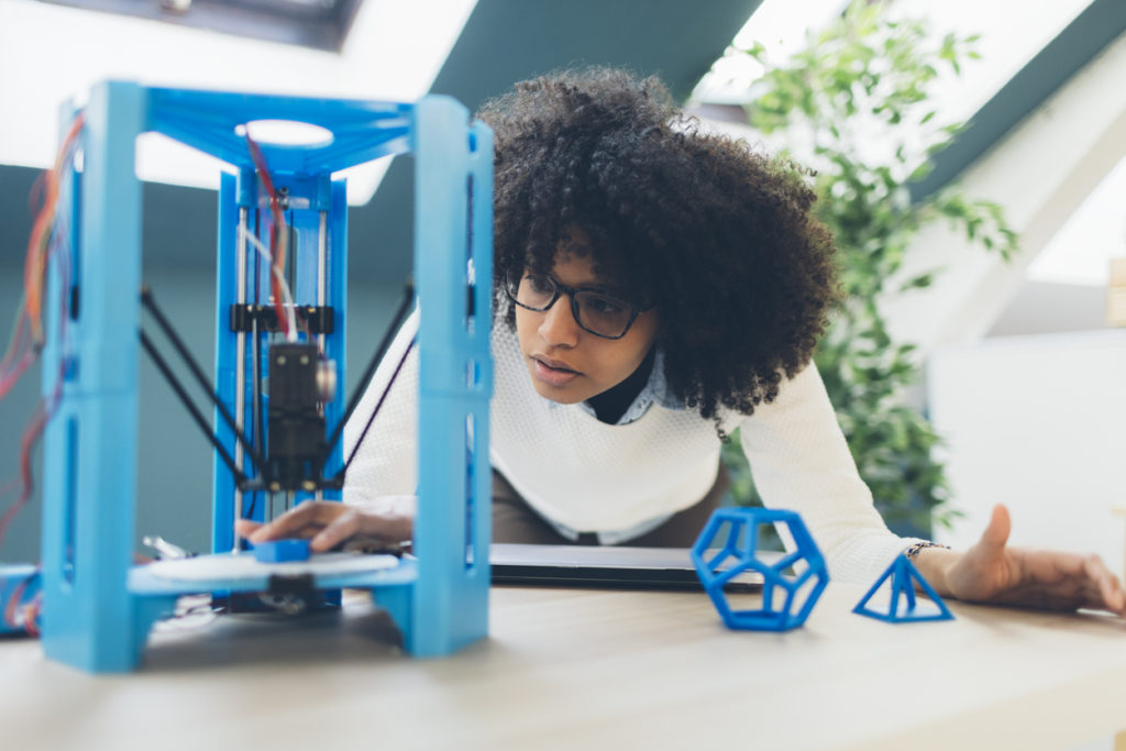
Though the concept of combining MRI imaging and robotic technology has been studied before (including work from this team on an MRI-Guided Prostate Therapy Robot now in clinical trials), this is the first time that both systems have successfully worked together. The team uses customized piezoelectric actuators and a custom-designed control system so that the robotic frame can use closed-loop motion sensing and control, making it more accurate than previous designs. It is faster, more intuitive, performs low-level position computation, and can adjust itself for better results. This adjustment addresses a previous challenge that surgeons faced when working with previous designs. Needing to manually adjust the robot for each precise movement was time-consuming and taxing for a surgeon. MRIs are also particularly harsh in the requirements that they impose on robotic systems due to their electromagnetic environments. Thus, the prototype is primarily made of machined and 3-D printed plastic, a nonconducting, nonmetallic and nonmagnetic material.

The robotic system is tested with an MRI.
“We’re thrilled about this neurorobot being used in future DBS procedures,” said Hao Su, graduate research student. “This prototype can treat certain mental diseases, so we’re excited about this design’s future use in hospitals.”
Find more articles about “Robotics” in IEEE Xplore.