In studies on artificial organs and organ-on-a-chip technologies, the heart has been one of the most difficult organs to simulate—until now. Researchers from the University of Alabama at Birmingham have developed biomimetic cardiac tissue chips (CTC)–cell culture models that can accurately mimic complex blood flow stresses associated with the heart’s pressure-volume changes. Researchers hope these tissue chips will speed drug and medical device research and increase safety by identifying harmful drugs before they reach human trials.
Researchers created the chips using three-dimensional (3D) cell fibers suspended in a microfluidic chamber between two posts and integrated within a flow loop, as shown in Figure 1. The fibers are subjected to hemodynamic loading, replicating the pressure-volume changes seen in the heart’s left ventricle. Various parameters associated with heart function, like heart rate, peak-systolic pressure, end-diastolic pressure and volume, end-systolic pressure and volume, and duration ratio between systolic and diastolic, can all be precisely manipulated. This allows the culture of cardiac cells under developmental, normal, and disease states.
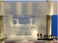
Figure 1: Image of the CTC prior to testing.
“The heart is a pump and the circulatory system is a network of fluidic channels perfused by the heart,” said Palaniappan Sethu, associate professor in the Department of Biomedical Engineering at the University of Alabama at Birmingham’s School of Engineering. “As engineers, we were able to bring a deeper understanding as to how to replicate this fluid flow in a lab setting.”
The CTC is unique because it can reproduce the stresses associated with pressure overload and volume overload, as show in Figure 2. Under pressure overload, the cells within the CTC see increased hypertrophic remodeling and fibrosis. Cells subjected to prolonged volume overload experience thinning and elongation. The new model builds on prior work to recreate 3D cardiac tissue by organizing heart cells, including cardiomyocytes, cardiac fibroblasts, and endothelial cells, in an aligned 3D tissue.
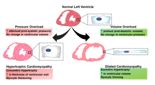
Figure 2: How pressure and volume changes impact heart cells.
A major reason for withdrawal of regulator-approved drugs is their unintended toxic effects on the heart, sometimes leading to fatal heart rhythm disorders. Because CTCs can provide a better understanding of disease progression and drug toxicity, researchers hope to detect these issues earlier on.
“Advances in cardiac cell cultures, such as CTCs, will allow researchers and regulators to screen drug candidates that may harm the heart before the drugs are tested on patients,” Sethu said.
The researchers are now working to integrate the CTCs with chips representative of other organs and tissue. Broader tissue chips would more accurately recreate human physiology for research and drug testing.
“We are just getting started with building physiologically relevant systems to model cardiac tissue in the heart,” Sethu said. He added that these systems will continually evolve in terms of functionality, throughput, accuracy and ease of use.
For more information on tissue engineering, visit the IEEE Xplore Digital Library.